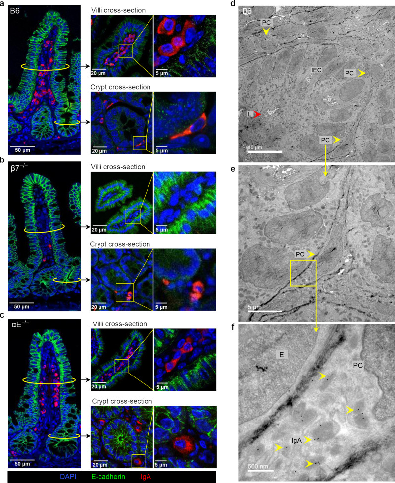
Fig. 5. A subset of IgA + ASC acquire and elongated/sickled morphology near the crypt base of B6 mice but not in αE- or β7-deficient mice.
a Round morphology of IgA + ASC in the mid-villous region, whereas a subset of IgA + ASC near the crypt base of B6 mice have an elongated (sickle-like) morphology (coronal and cross sections at indicated levels). b Absence of IgA + ASC in the mid-villous and near crypt base regions in β7−/− mice. c Round morphology of IgA + ASC in the mid-villous region and around the crypt base of αE−/− mice. Increasing magnification of the villous crypt base of B6 mice (d) Three cells (PC) with sickled morphology in contact with IEC (yellow arrowheads) (e) Higher magnification of an adherent cell with sickled morphology (f) Direct cell to cell contact between IEC and sickled cell with extensive RER and IgA immunogold particles (representative TEM images, L = lumen, E = epithelial cell).

