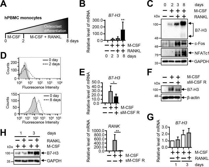
Fig. 1. B7–H3 expression is increased during differentiation of human osteoclasts.
A A schematic diagram outlining the process of osteoclast differentiation from human PBMCs. B B7–H3 mRNA expression during human osteoclast differentiation measured using RT-qPCR. C Whole-cell lysates were immunoblotted with B7–H3, c-Fos, and NFATc1 Abs. D At days 0, 2, and 8, the cell-surface B7–H3 expression was assessed by flow cytometry (solid line: monocytes; dashed line: OCPs or mature osteoclasts; gray shaded: isotype control). E, F OCPs were cultured as described in Fig. 1A. OCPs were incubated with M-CSF (20 ng/ml) in the presence or absence of recombinant human M-CSF receptor/Fc chimera (sM-CSF R) overnight. The expression of B7–H3 and RANK mRNA was measured using RT-qPCR and the expression of B7–H3 protein was detected using Western blot. G, H OCPs were cultured with M-CSF (20 ng/ml) in the presence of RANKL (40 ng/ml) for one or three days. RT-qPCR and Western blot were performed to detect the expression of B7–H3. (B, E, G) mRNA levels were normalized with GAPDH expression.

