
Fig. 5.
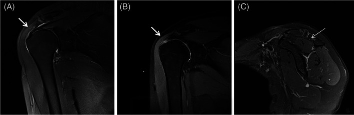
Images of a 47‐year‐old woman undergoing arthroscopic debridement before and 18 months after operation. (A) MRI before operation, showing high intensity signal on the bursal side of the supraspinatus tendon (arrow). (B) MRI at 18 months postoperatively, showing insufficient thickness without discontinuity in the supraspinatus tendon (Sugaya grade III). (C) MRI shows moderate atrophy (grade II) of the supraspinatus muscles without fat infiltration (grade 0) at 18‐month follow‐up.