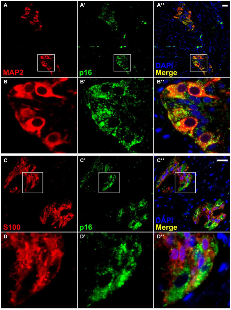
FIGURE 5.
Co-localization of p16 with a neuronal marker in the myenteric plexus, but with little or no co-localization with a glial cell marker. Immunofluorescence for a neuronal marker, MAP2 (A,B), shows co-staining with p16 (A′,A″,B′,B″). However, immunofluorescence for a glial cell marker, S100 (C,D), shows minimal co-localization with p16 (C′,C″,D′,D″). It is noteworthy that p16 staining is present mainly in the neuronal cytoplasm and not the nucleus. Most nuclear staining observed was peripheral, and it was impossible to rule out overlap with the cytoplasm. Neuronal nuclei are present but do not take up DAPI stain as well as other cell types. S100 staining was not further quantified. Scale bar represents 25 μm.

