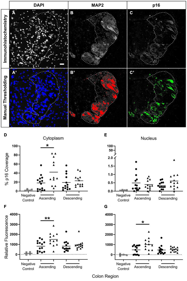
FIGURE 6.
Increased p16 protein expression with advanced age in neuronal cytoplasm in ascending but not descending colon. Thresholding was used to identify positive immunostaining. Panels (A–C) show the original immunostaining, and panels (A′,B′,C′) show the respective thresholding, with areas of positive staining shown in color. Images DAPI (A,A′) and MAP2 (B,B′) determined positive and negative areas of staining. For p16 (C,C′) the level of fluorescence in the surrounding smooth muscle was measured, and the threshold was set at twice this background level. Scale bar in A is 25 μm. The results were calculated using two methods. The first, is a calculation of the percentage of area covered by p16-positive staining in the region of interest [shown in panels (D,E)] as determined by thresholding. The second method is a measurement of the average level of p16 fluorescence in the region of interest relative to background (F,G). Results are for the cytoplasm (D,F) and the nucleus (E,G). Bars indicate data mean. N = 52: adult ascending colon n = 14, elderly ascending n = 12, adult descending n = 13, elderly descending n = 13. *p < 0.05 and **p < 0.01. Student’s t test used to compare between age groups. Panel (D) shows a significant increase in p16 coverage in the neuronal cytoplasm of the ascending colon when comparing samples from adult (25–60 years old; •) and the elderly (70+ years old; ▲). Panel (E) shows no age-related changes in p16 coverage in neuronal nuclei for either colon region. Panels (F,G) show age-related increases in p16 fluorescence in the ascending colon in cytoplasm (F) and nuclei (G) of myenteric plexus neurons; changes in the nuclei may be due to overlap with the cytoplasm.

