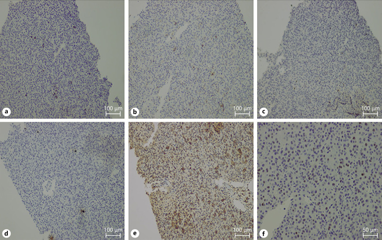
Fig. 2.
Immunostaining using a needle biopsy specimen of sacral metastases. CD3+ (a), CD4+, and CD8+ cells were observed. CD4+ cells were predominant in the TILs (b, c). We also found the expression of PD-1 in 10% of TILs (d), along with a dense expression of PD-L1 in the tumor (e). (f) A considerable number of Foxp3+ TILs were also observed in HCC tissues. The antibodies used were CD3 and CD4 (Roche Diagnostics, Tokyo, Japan), CD8 (Nichirei Bioscience, Tokyo, Japan), PD-1, PD-L1, and FoxP3 (Abcam, Cambridge, UK). CD3 staining (a), CD4 staining (b), CD8 staining (c), PD-1 staining (d), PD-L1 staining (e), and FOXP3 staining (f). Bar, ×200. TILs, tumor infiltrated lymphocytes; PD-1, programmed cell death 1; PD-L1, programmed cell death ligand 1; HCC, hepatocellular carcinoma.

