
Figure 6: Individual values for cerebrospinal fluid (CSF) levels of (A) MHPG as a function of HVA, (B) HVA as a function of the putamen/occipital (PUT/OCC) ratio of 18F-DOPA-derived radioactivity, (C) HVA as a function of the washout percent of 18F-DOPA-derived radioactivity, and (D) MHPG as a function of septal myocardial 18F-dopamine- (18F-DA-) derived radioactivity in synucleinopathies and control subjects.
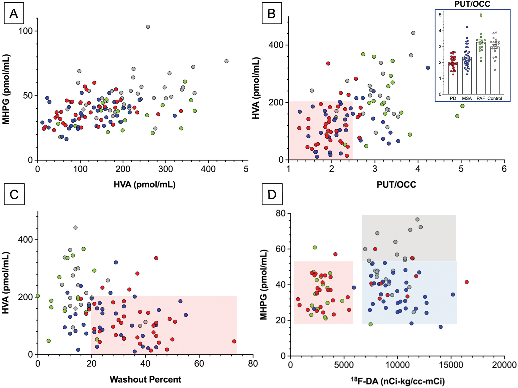
Abbreviations: HVA=homovanillic acid; MSA=multiple system atrophy (total N=37, blue); PAF=pure autonomic failure (total N=19). PD=Parkinson disease (Total N=36). PD data are in red, multiple system atrophy, MSA blue, PAF green, and control subjects gray (Total N=24). Inset in (B) shows individual PUT/OCC ratios, with means ± SEM. Pink rectangles in (B) and (C) placed visually to emphasize low HVA, PUT/OCC ratios, and increased 18F-DOPA washout precents in PD and MSA. In (D), gray rectangle placed visually to depict normal values. The pink and blue rectangles in (D) are placed visually to indicate low 18F-DA-derived radioactivity in PD and PAF and normal radioactivity in MSA.