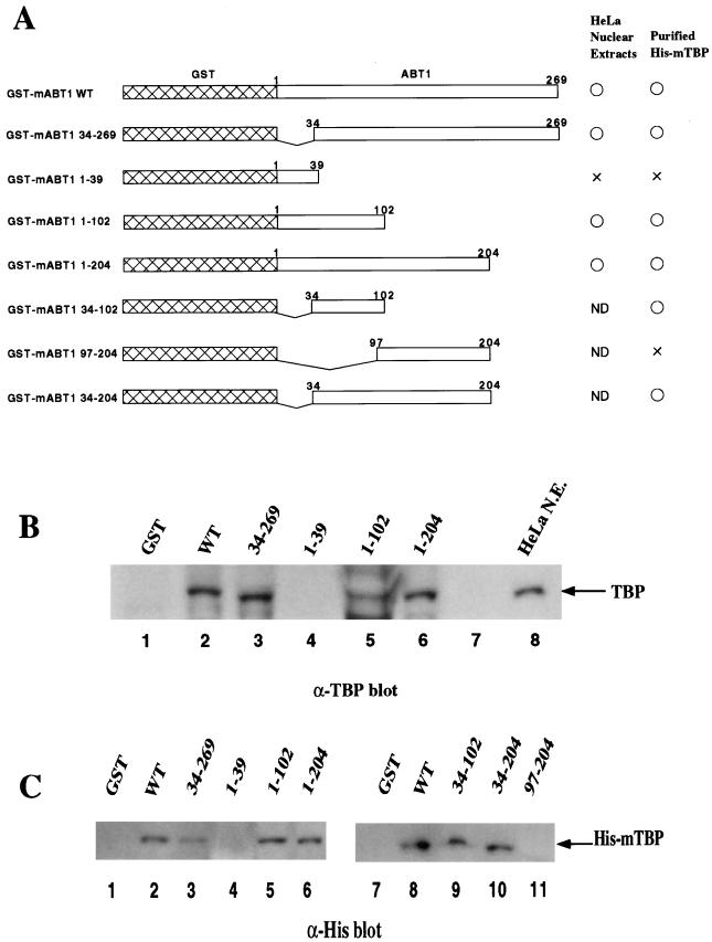
FIG. 5.
Delineation of the TBP-binding region of mABT1. (A) Schematic representation of a series of GST-mABT1 mutants and summary of the binding characteristics. Numbers show the sequence position of amino acids in mABT1. Results of the interaction analysis with TBP in HeLa nuclear extracts and purified His-mTBP are summarized to the right (○, interaction; ×, no interaction; ND, not determined). The binding and washing conditions for TBP binding to the GST-mABT1 proteins were the same as in Fig. 4C. (B) Interaction of the GST-mABT1 deletion mutants with TBP in HeLa nuclear extracts. The series of GST-mABT1 deletion mutants were incubated with HeLa nuclear extracts and washed with PBS containing Triton X-100. Proteins bound to the GST fusion proteins were then detected by immunoblotting with an anti-TBP antibody. Lane 1, GST; lane 2, mABT1 WT; lane 3; mABT1(34-269); lane 4, mABT1(1-39); lane 5, mABT1(1-102); lane 6, mABT1(1-204); lane 8, control HeLa nuclear extract (N.E.) (C) The series of GST-mABT1 deletion mutants were incubated with His-mTBP and washed with PBS containing 0.1% Triton X-100. Proteins bound to the GST fusion proteins were detected by immunoblotting with an anti-His antibody. Lanes 1 and 7, GST; lanes 2 and 8, mABT1 WT; lane 3, mABT1(34-269); lane 4, mABT1(1-39); lane 5, mABT1(1-102); lane 6, mABT1(1-204); lane 9, mABT1(34-102); lane 10, mABT1(34-204); lane 11, mABT1(97-204).

