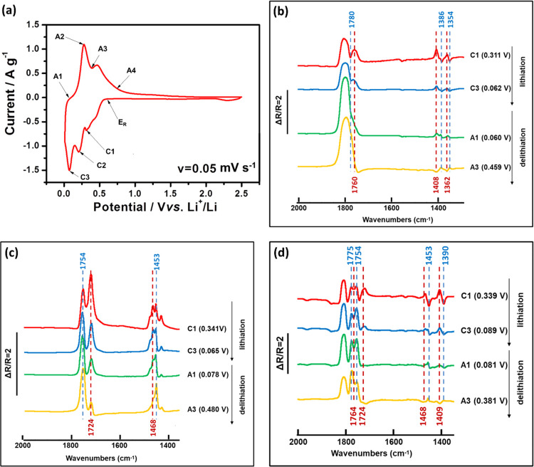
Figure 7.
(a) First cycle CV of the Si electrode tested in the three-electrode in situ MFTIRS cell in 1 M LiPF6/EC-DMC (scan rate v = 0.05 mV s–1). The in situ MFTIR spectra of the Si electrode in (b) 1 M LiPF6/PC (ER = 0.611 V), (c) 1 M LiPF6/DMC (ER = 0.590 V), and (d) 1 M LiPF6/EC-DMC (ER = 0.671 V) at C1, C3, A1, and A3 points during the first CV cycle.

