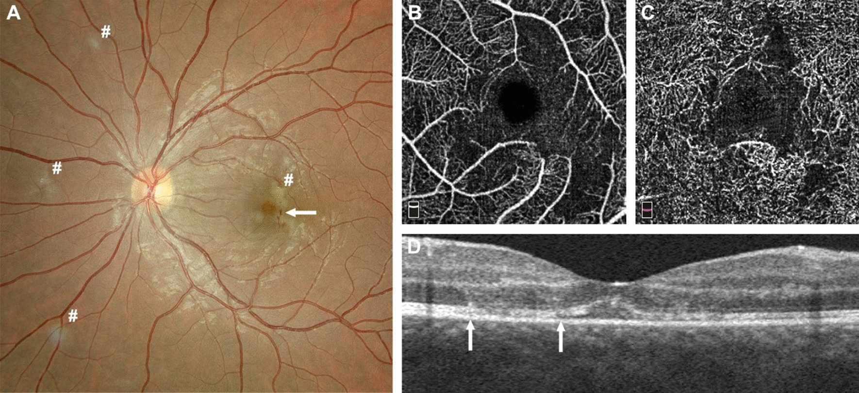
Fig. 2.

A Fundus photography of the left eye: presence of hemorrhages in the posterior pole (arrow), zones of focal ischemia vaso-occlusive retinitis also present nasally and around the macula (# in the picture). B–D Optical coherence tomography angiography (OCTA, Optovue imaging) revealing alterations of the superficial plexus of the retina (B) and of the deep retinal plexus (C); note the disappearance of the vessels due to occlusive vasculitis. D Horizontal optical coherence tomography (OCT, Optovue imaging) revealing alterations of the retinal layers (between the arrows) due to local capillary ischemia
