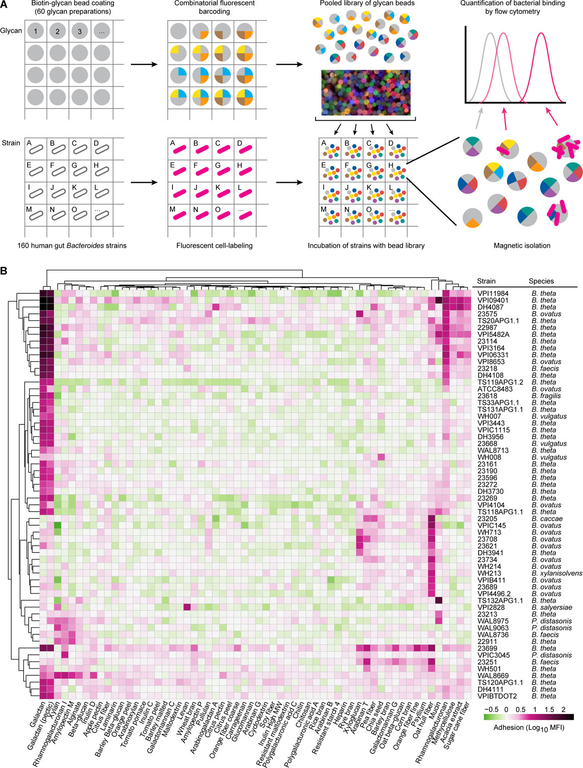
Figure 1: Multiplex screening of human gut Bacteroides and Parabacteroides species/strains for their ability to bind to fluorescent, barcoded, paramagnetic microscopic glycan-coated beads.

(A) Schematic design of how the polysaccharide bead library was generated and used to simultaneously characterize the glycan binding phenotypes of multiple bacterial strains. The matrix depicts how a given bacterial strain was added to a given well of a multi-well plate, labeled with Syto-82 and incubated with the 60-member library of fluorescently barcoded beads (depicted as a cartoon as well as by a photograph of labeled beads). Beads were recovered from each monoculture based on their magnetism and then subjected to flow cytometry according to the gating strategy depicted in Table S1C to identify bead types with bound bacteria. (B) Quantification of bacterial adhesion for all strains that exhibited glycan-dependent binding (>4-fold above the average level of fluorescence of “empty” beads not coated with glycans). The log10 geometric mean fluorescent intensity in the Syto-82 channel for each population of beads in each well relative to empty beads is indicated by the color bar. Columns denote the carbohydrate preparation coated on each bead type. Bacterial strains incubated with beads are listed in the rows along with their species classification. Binding profiles for a given bead type and strain are hierarchically clustered using Ward’s minimum variance (Ward, 1963). See also Table S1, S2.
