
Fig. 3. Tslpr-deficient mice have increased MRSA burden during in vivo skin infection.
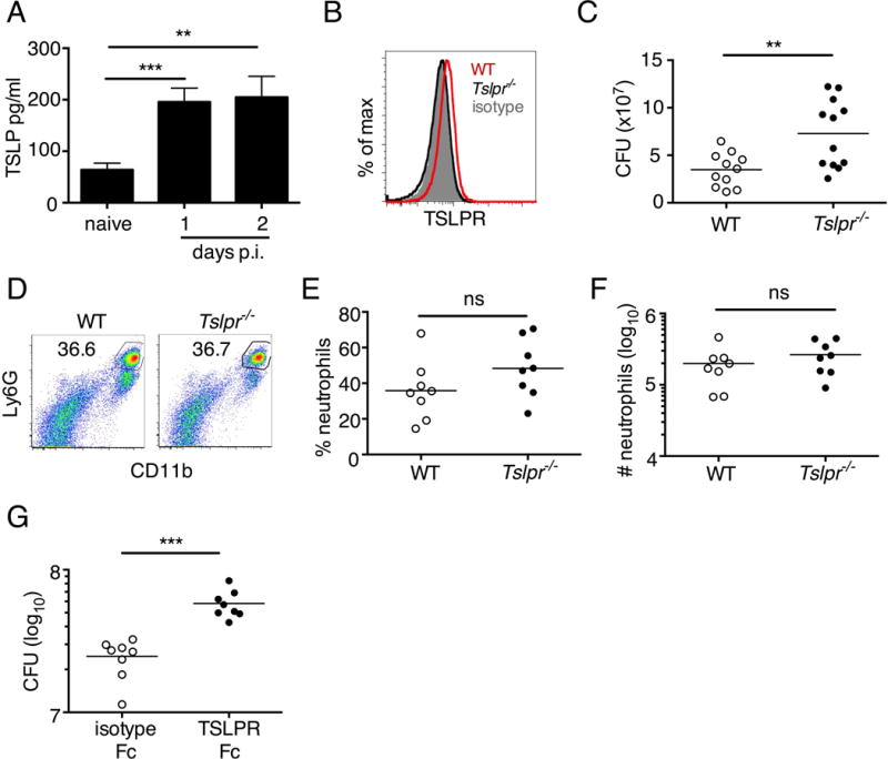
(A–G) Mice were infected with MRSA i.d. in the ear. (A) TSLP protein expression in the ear after i.d. MRSA infection (n=4), naïve controls were mock-infected with PBS only. (B–G) Ears were analyzed on day 1 p.i. (B) Representative TSLPR expression on neutrophils. (C) Analysis of CFU in the ear of WT (n=11) and Tslpr−/− mice (n=12 ears). (D–F) Analysis of neutrophils in the ear. Shown are representative FACS plots (D) and percent (E) and total number (F) of neutrophils from WT and Tslpr−/− mice (n=8 ears). (G) CFU of MRSA in the ear on day 2 post-i.d. ear infection of WT mice treated with human IgG1 Fc isotype control or TSLPR Fc (two-tailed Mann-Whitney test) (n=8 ears). *, p < .05; **, p < .01; ***, p < .001; ns = not significant. a–f, two-tailed Student’s t-test. Data are representative of 3 (or 2 for (G)) independent experiments.