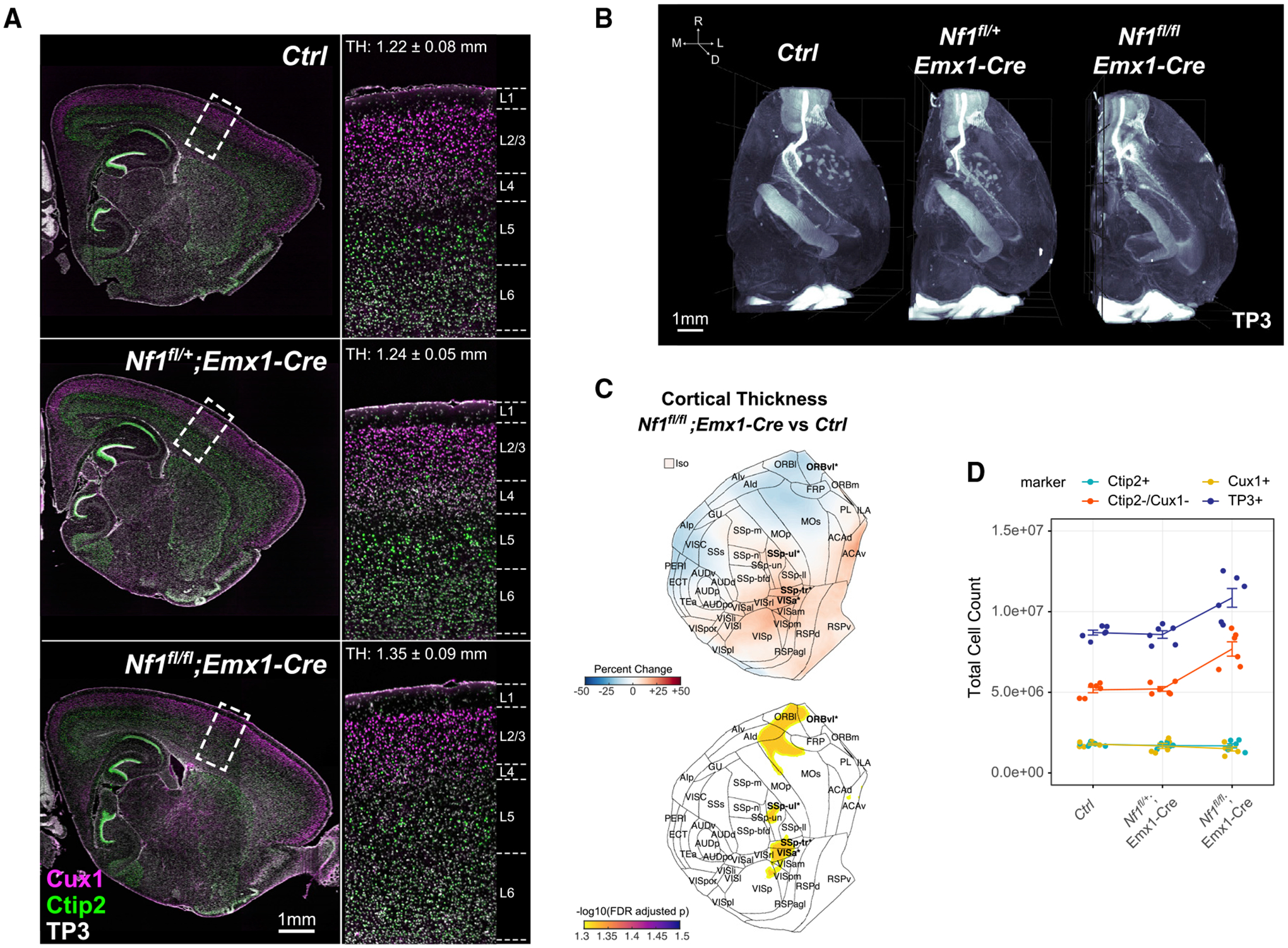
Figure 5. Nf1 deletion induces cortical thickening driven by increased numbers of non-excitatory neuronal cell types.

(A) Optical sagittal sections of immunolabeled lower layer (Ctip2+) and upper layer (Cux1+) neurons in P14 Ctrl, Nf1fl/+;Emx1-Cre, and Nf1fl/fl;Emx1-Cre brain hemispheres. Zoomed-in regions of boxed cortical areas near the somatosensory cortex showing expected localization of upper and lower layer neurons. Average cortical thickness (TH) measurements indicated for full 3D somatosensory volumes.
(B) Three-dimensional rendering of cell nuclei in Ctrl, Nf1fl/+;Emx1-Cre, and Nf1fl/fl;Emx1-Cre brain hemispheres.
(C) Flattened isocortex displaying percentage change and FDR-adjusted p values for cortical thickness in Nf1fl/fl;Emx1-Cre across 43 cortical regions and the full isocortex (Iso) compared with Ctrl. Significant regions are bolded (FDR < 0.05) and starred.
(D) Total isocortex counts of each cell-type class measured across all Nf1+/+, Nf1fl/+;Emx1-Cre, and Nf1fl/fl;Emx1-Cre samples (n = 6, mean ± SEM).
