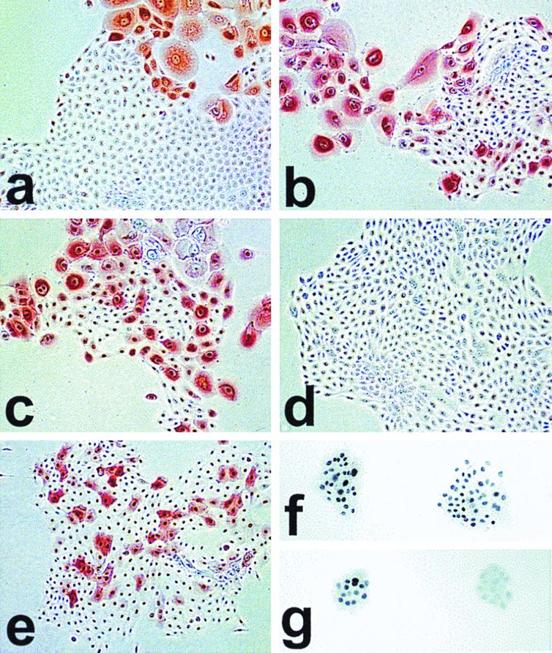
FIG. 4.

Immunocytochemical staining of cell lines for p16INK4a (a to e, phase contrast) and p53 (f and g, bright field). Cells were plated at clonal density in K-sfm and cultured for 4 to 7 days before staining. Life span designations are as for Fig. 3a. (a) N, 28 PD (mid-life span); (b) N/BABE-2G, 55 PD (near senescence); (c) N/TERT-2F, 80 PD (SGP); (d) N/TERT-1, 114 PD (RDI+43); (e) LiF-Ep/TERT-1, 67 PD (RDI+28); (f) LiF-Ep/TERT-1, 39 PD (early RDI); (g) LiF-Ep/TERT-1, 48 PD. Note the heterogeneity of p16 expression in normal N cells in panels a and b, confined to large nondividing cells, with small proliferating cells expressing no detectable p16 whether at mid-life span or in rare dividing cells in the nearly senescent population. As shown in panel c, hTERT-transduced keratinocytes continue the normal pattern of p16 expression during SGP. Some RDI lines that emerge from SGP retain the normal pattern of p16 expression, while some cease expressing p16. The p53+ colonies in panel f are representative of >99% of colonies in the early RDI LiF-Ep/TERT-1 population (as confirmed by the Western blot shown in Fig. 3b). In panel g, the p53+ colony at the left and the p53− colony at the right are representative of the mixed nature of the LiF-Ep/TERT-1 RDI population three passages (9 PD) later.
