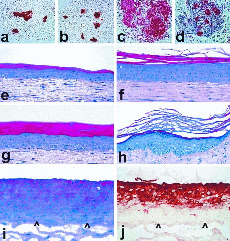
FIG. 6.
Differentiation-related protein expression and epithelial tissue formation by hTERT-immortalized keratinocytes. Cells were cultured on conventional plastic dishes for 7 to 9 days in K-sfm (a and b) or in the feeder cell-FAD system (c and d) or were seeded on collagen gels to generate organotypic cultures (e to j), one of which was then grafted to the skin of an athymic mouse (h). Panels a (LiF-Ep, 28 PD) and b (LiF-Ep/TERT-1, 66 PD [RDI+43]) were immunostained for the suprabasal epidermal differentiation keratin K10. Panels c (OKF6, 30 PD) and d (OKF6/TERT-2, 122 PD [RDI+63]) were immunostained for the keratinocyte terminal differentiation protein involucrin. (e) N, 37 PD (mid-life span); f, N/TERT-1, 91 PD (RDI+24); g, LiF-Ep/TERT-1, 90 PD (RDI+55); h, a replicate organotypic culture of the one shown in panel g, 42 days after transplantation as a skin graft to an athymic mouse; i and j, OKF6/TERT-2, 86 PD (RDI+47). Panels e to i were stained with H&E (note that the intense blue of the cell layers below the stratum corneum in the epithelium shown in panel h is the result of a slightly different staining formulation and has no biological significance). Panel j was immunostained for keratin K13. Arrowheads point to the basal layer of the organotypic epithelium, showing that K13 expression begins several cell layers above the first suprabasal layer.

