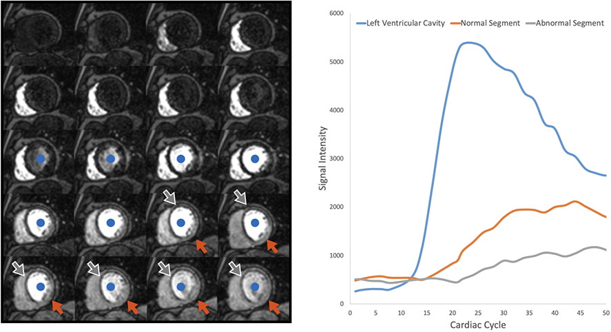
Figure 3. First pass perfusion.
Left Panel: A single mid-ventricular short axis slice acquired repetitively as contrast first enters the right ventricle, then the left ventricle (blue dot), and finally perfuses the myocardium. More contrast enters the non-ischemic segments of the myocardium (orange arrow) when compared to the ischemic segments (grey arrow). Time intensity curves representing the left ventricular cavity, an ischemic segment, and a normal segment are shown.

