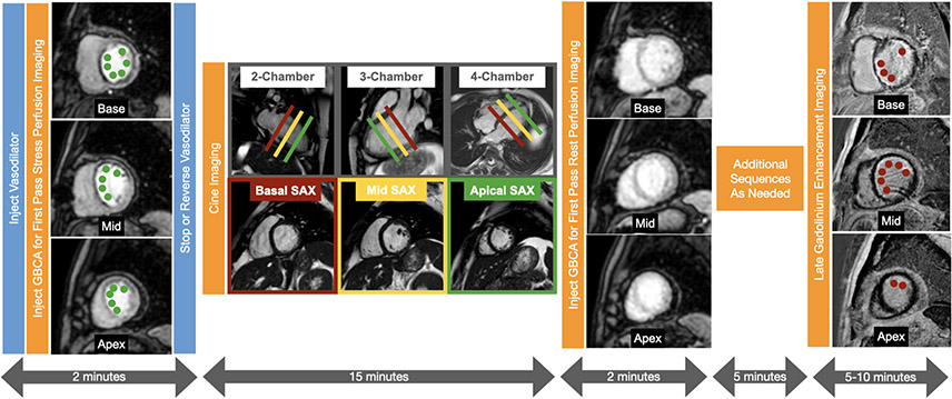
Figure 4. Typical vasodilator stress cardiac magnetic resonance protocol.
First pass perfusion images using a gadolinium-based contrast agent (GBCA) are acquired in 3 short axis slices of the left ventricle during hyperemic conditions to assess for ischemia. During the next 15 minutes, cine images are acquired in the multiple short axis and long axis views to assess cardiac anatomy and function. Next first pass perfusion images are acquired in 3 short axis slices during resting conditions. After a 5 minute delay, late gadolinium enhancement images are acquired in multiple short axis and long axis views to assess for viability. Large perfusion defects involving most of the basal slice and the mid to apical septal, anterior, and anterolateral segments are present (adjacent to the green dots) on the stress perfusion images but not on the rest perfusion images which are normal. The perfusion defect extends beyond the thin rim of late gadolinium enhancement (red dots) consistent with subendocardial myocardial infarction with significant peri-infarction ischemia.

