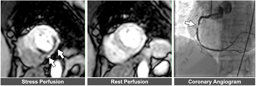
Figure 5. Example of abnormal stress CMR perfusion.
Left panel: Perfusion defect in mid-ventricular short axis slice denoted by arrows. . Middle panel Normal perfusion noted in same slice position during resting conditions. Right panel: Severe right coronary artery stenosis corresponding to perfusion defect.

