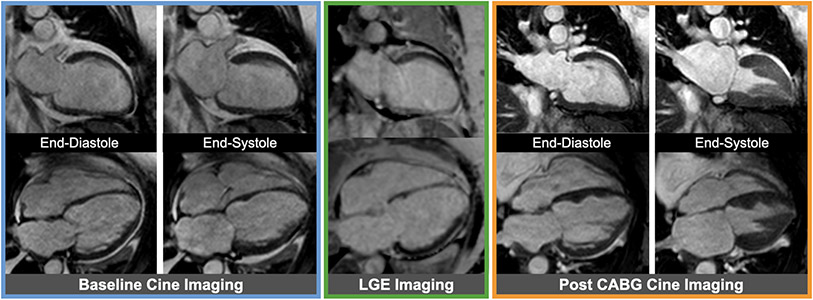
Figure 8. Example of viable myocardium.
Baseline cine images (blue box) from a patient with severe multi-vessel coronary artery disease showing severely reduced ejection fraction. Late gadolinium enhancement (LGE) images (green lox) without any significant LGE representing fully viable myocardium are shown in the same cardiac views; of note, the mid to apical septum in the 4-chamber view has mildly increased signal intensity not sufficient enough to be classified as myocardial infarction. Corresponding cine images (orange box) showing improved ejection fraction following coronary artery bypass grafting (CABG).

