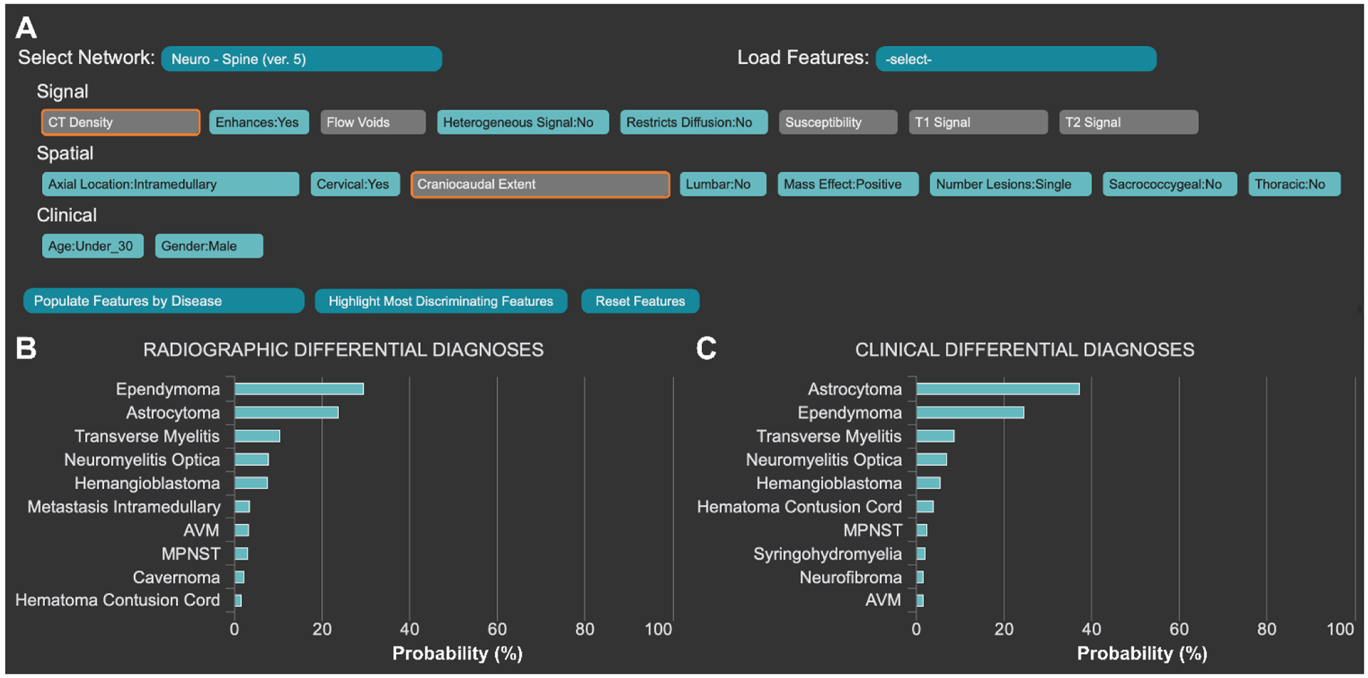
Figure 2.

The Adaptive Radiology Interpretation and Education System (ARIES) distinguishes between ependymoma and astrocytoma in the spine network. (A) Features based on signal, spatial and clinical information are selected by the trainee in blue. Unselected features are grey and the most differentiating unanswered features are highlighted in orange. Differential diagnoses by (B) imaging features only vs (C) a combination of clinical and imaging features are derived from manually selected features (A). Probabilities are calculated by a naïve Bayes network.
