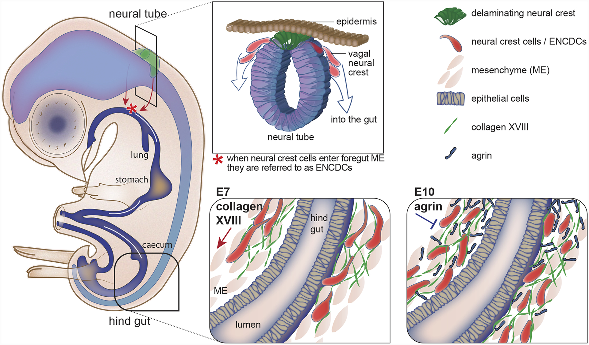
Figure 1. Neural crest cells secrete matrix components to control their own migration.

Most of the enteric nervous system of the gut is derived from vagal neural crest cells that undergo an epithelial-to-mesenchymal transition (EMT) at the dorsal neural tube, delaminate, and then migrate and colonize the mesenchyme of the foregut. Once in the gut, the neural crest cells are referred to as enteric neural crest-derived cells (ENCDCs) and they undergo a long migration along the basement membrane (BM) of gut blood vessel (not shown) to populate the entire gut. At embryonic day 7 (E7) the wave front of the ENCDCs reaches the distal hind gut where they secrete collagen XVIII, which promotes ENCDC migration. Later, at E10, the ENCDCs secrete agrin, which inhibits ENCDC migration.
