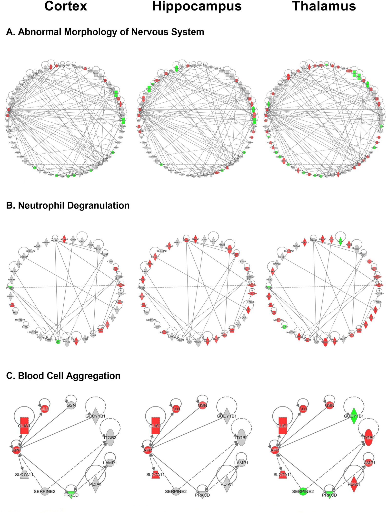
Figure 4. IPA downstream effects analysis networks.

A. Abnormal morphology of nervous system network for the cortex, hippocampus and thalamus of rTg-DI rats including all associated differentially expressed proteins (≥ 50% increase or ≥ 34% decrease, p < 0.05). B. Neutrophil degranulation network depicted for the cortex, hippocampus and thalamus of rTg-DI rats. C. Aggregation of blood cells network created in IPA depicted for the cortex, hippocampus, and thalamus of rTg-DI rats. Red shading indicates increased, green decreased and grey not differentially expressed proteins in that region.
