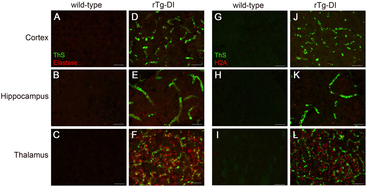
Figure 7. Increased immunolabeling for neutrophil elastase and H2A exclusively in the thalamus of rTg-DI rats.

Brain sections from the present cohort of 12 M wild-type rats (A,B,C and G,H,I) and rTg-DI rats (D,E,F and J,K,L) were stained with thioflavin S to detect microvascular fibrillar amyloid (green) and rabbit polyclonal antibody to neutrophil elastase (red) (A-F), or rabbit polyclonal antibody to histone 2A (red) (G-L). Scale bars = 50 μm. Representative images show increased neutrophil elastase and histone 2A is increased only in the thalamus of rTg-DI rats.
