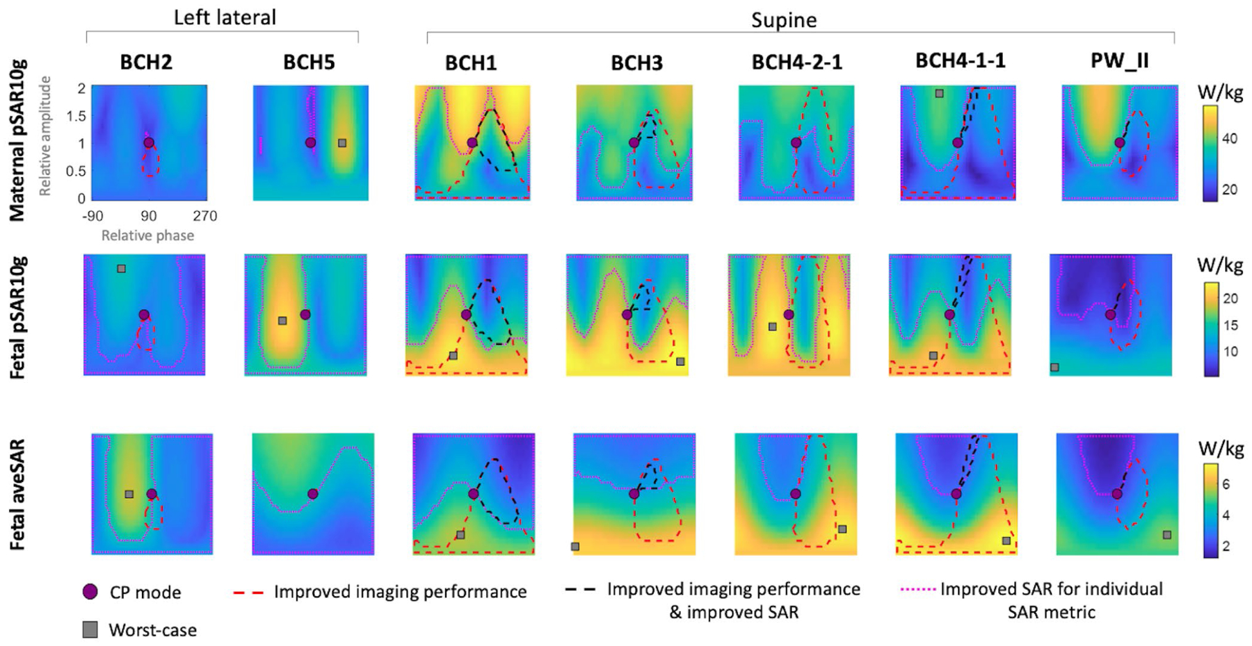
FIGURE 3.

Maternal and fetal peak local SAR (pSAR10g) and fetal average SAR (aveSAR) for different RF shim settings where the relative amplitude and relative phase of the two channels are varied from 0 to 2 (vertical axis) and −90° to 270° (horizontal axis), respectively. All shim settings are normalized to a maternal whole-body average SAR (wbSAR) of 2 W/kg. CP mode: circularly polarized birdcage mode, improved imaging performance: both average and CV are improved compared with CP mode, improved SAR: all three SAR metrics (maternal pSAR10g, fetal pSAR10g, and fetal aveSAR) are less than the CP mode values of the corresponding body model, improved SAR for individual SAR metric: the individual SAR metric in each subplot is lower than its corresponding CP mode value, worst-case: RF shim setting that maximizes an individual SAR metric. For some cases, the worst-case shim setting has a relative amplitude >2; hence, it is not shown on the plot
