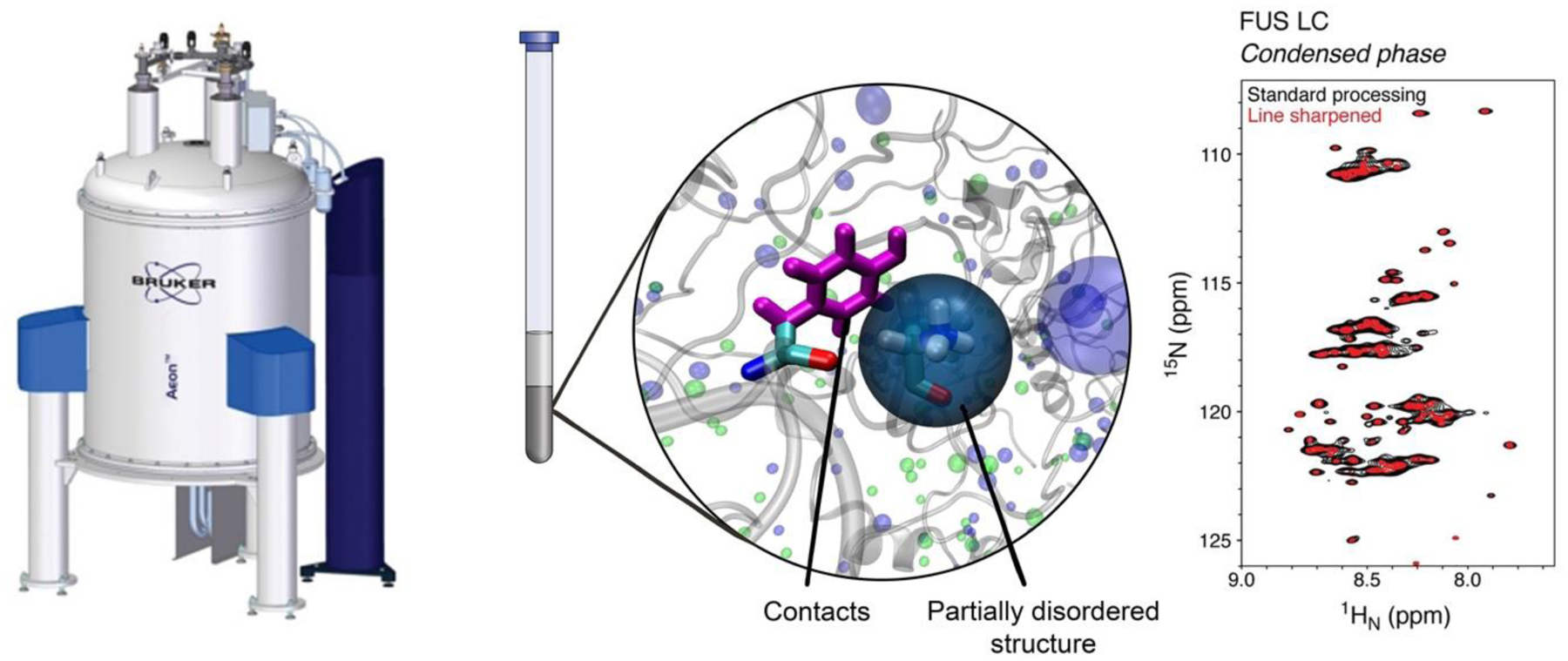
Figure 2. NMR spectroscopy of phase separation.

NMR spectroscopy of samples where the condensed phase (gray, center) fills the observation (coil) volume enables direct interrogation of structure and disorder in proteins and their contacts (center) with residue-by-residue resolution (right). Image concept by Vinald Francis.
