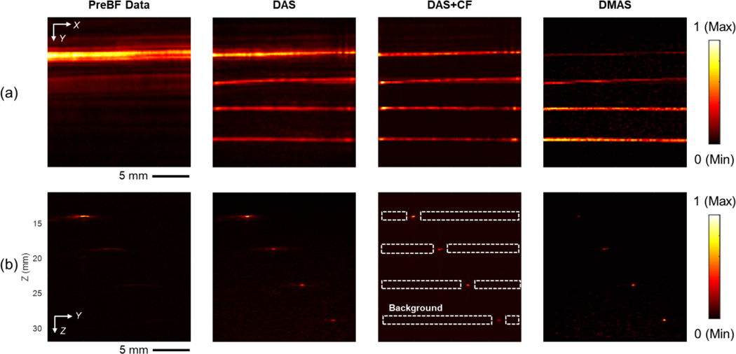
Fig. 6.
The PA imaging results of the pencil-lead phantom, four pencil leads were placed at 14 mm, 19 mm, 24 mm, and 29 mm from the surface of the ultrasound transducer, which is 1.6 mm, 6.6 mm, 11.6 mm, and 16.6 mm away from the focal depth of the transducer, respectively. (a) The MIP images of pre-beamformed data, DAS, DAS+CF, and DMAS reconstruction. (b) The corresponding cross-section slices at X = 10 mm position. The depth axis unit presents the distance from the transducer surface (the focal depth is 12.4 mm). The color bar represents the normalized PA intensity. The dot section is the background used to compute SNR.

