Figure 3.
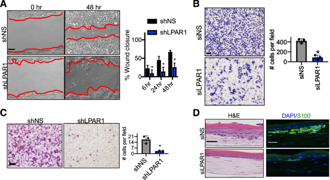
LPAR1 drives melanoma motility and invasiveness. A, A wound healing assay was performed in WM9 cells with the knockdown of LPAR1. Red lines indicate the leading edges of migrating cells. Scale bar, 50 μm. The percentage of wound closure after different time periods of migration was calculated and is shown. Error bars, SD; n = 3; *, P < 0.05. B, Transwell migration assay (without Matrigel coating) was performed for WM9 cells infected with luciferase shRNA (shLuc) or shLPAR1. Scale bar, 100 μm. Right, the number of migrated cells were quantified. Error bars, SD; n = 3; *, P < 0.05. C,Transwell invasion assay (with Matrigel coating) was performed in 1205Lu cells infected with shLuc or shLPAR1. Scale bar, 100 μm. The numbers of invaded cells were quantified. Error bars, SD; n = 3; *, P < 0.05. D, 1205Lu cells transfected with siNS or siLPAR1 were used to make skin reconstructs. S100, green; DAPI, blue. Scale bar for hematoxylin and eosin (H&E) staining, 50 μm. Scale bar for immunofluorescence staining, 20 μm.

