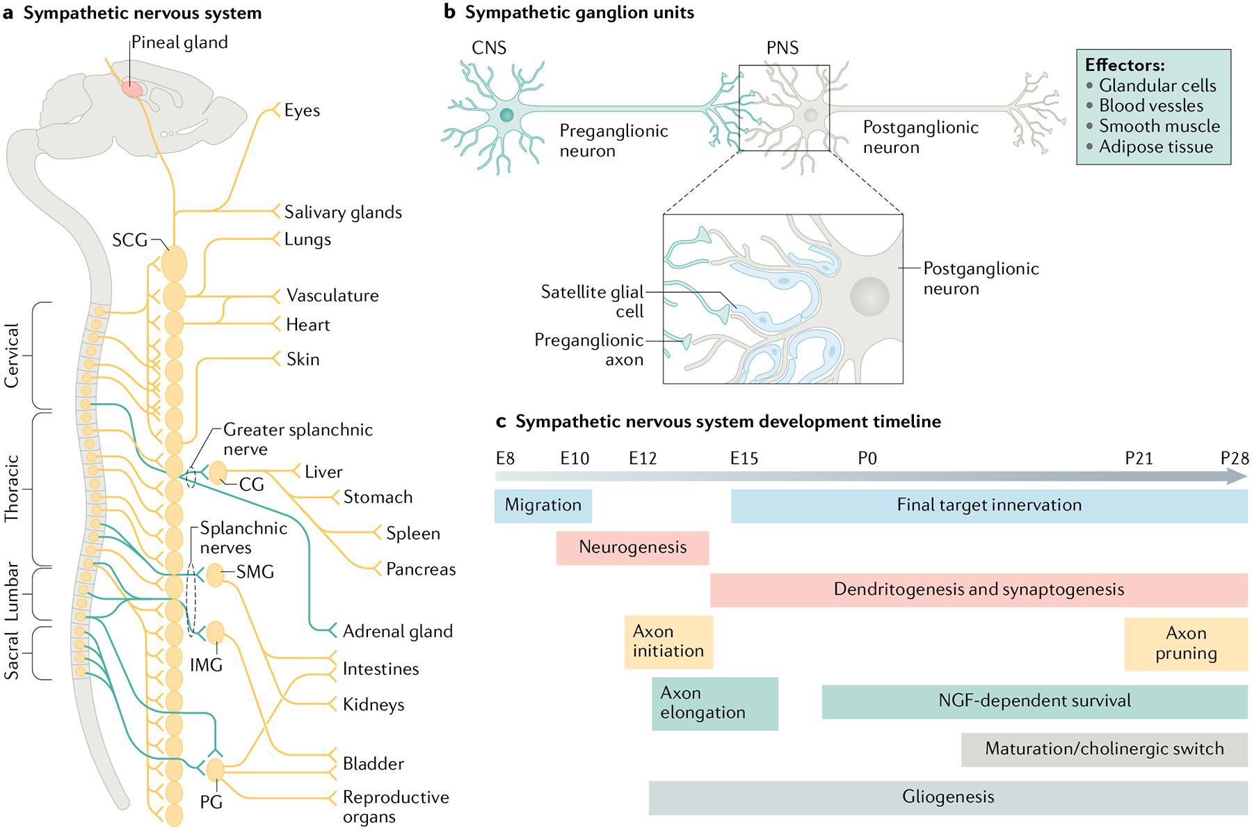
Fig. 1 |. Organization of the mouse sympathetic nervous system and timeline of developmental events.

a | Sympathetic neuron cell bodies located in either paravertebral (for example, superior cervical ganglia (SCG)) or prevertebral sympathetic ganglia (for example, celiac ganglia (CG), superior mesenteric ganglia (SMG), inferior mesenteric ganglia (IMG), or pelvic ganglia (PG)) send axonal projections during development to innervate diverse organs and/or tissues in the periphery. b | Postganglionic sympathetic neurons serve as a bridge between the central nervous system (CNS) and diverse peripheral organs through inter-neuronal synapses with preganglionic sympathetic neurons whose cell bodies lie in the spinal cord. Sympathetic ganglia also contain satellite glia cells that enwrap cell bodies, dendrites and synapses of postganglionic neurons. c | Key events in the development of postganglionic sympathetic neurons are correlated with axon innervation of peripheral organs, preganglionic input and gliogenesis. The embryonic (E) and postnatal (P) time-points are based on studies done in mice and rats. NGF, nerve growth factor; PNS, peripheral nervous system. Part a adapted with permission from REF.6, AAAS. Part c adapted with permission from REF.2, Annual Reviews.
