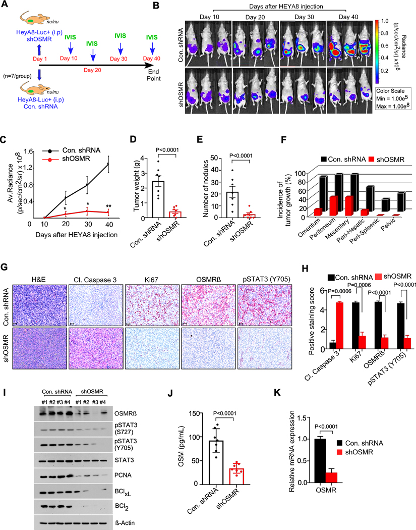
Figure. 3. OSMR knockdown reduced the growth and seeding of ovarian cancer cells in vivo.
A-C, Athymic female nude mice were injected with shControl-HEYA8-Luc+ cells intraperitoneally or shOSMR#1-HEYA8-Luc+ cells (n=7/group) and images were taken at the indicated days using IVIS100 bioluminescence imager. D-F, Mice from (b) were sacrificed at the end point (40th day) and total tumor weight, number of tumor nodules and the incidence of tumor growth were recorded. G-H, IHC of tumor tissues from (B) were performed using the antibodies indicated, then photographed and the antibody staining intensity was scored and quantitated. Scale bar, 50 μm. I, Three different tumor tissue lysates from each group in (B) were immunoblotted. J, Serum from the mice in (A) were collected (n=7) and ELISA was performed to determine OSM levels. K, qPCR showing relative mRNA expression of OSMR in tumor tissues of mice from (b). Data represent means ± SEM and Student’s t test (two tailed, unpaired) was performed to determine p-Value in (C, D, E, H, J, K).

