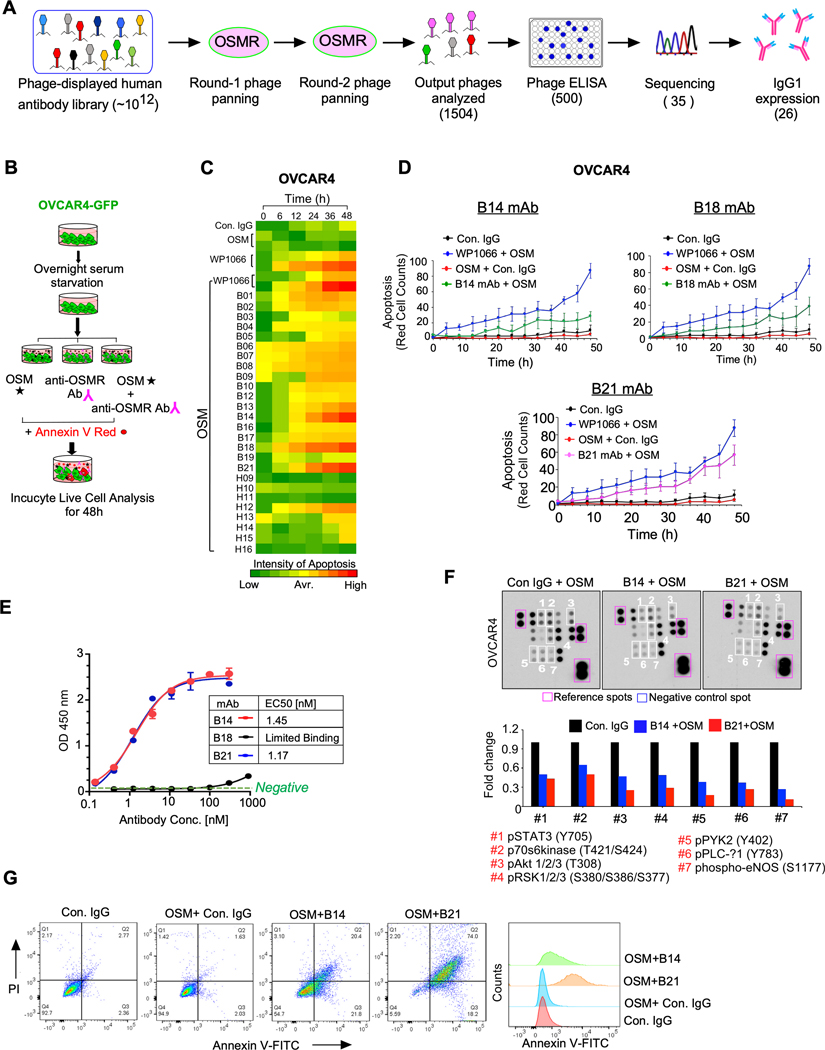
Figure. 4. Monoclonal antibody (mAb) of OSMR abrogates OSM-mediated oncogenic characteristics by blocking dimerization of OSMR.
A, Flowcharts demonstrate the scFv phage library panning and antibody selection process employed. B, Schema shows the flowchart of screening 26 anti-OSMR mAb by the ability to promote apoptosis in the presence of recombinant OSM (100ng/mL) in OVCAR4-GFP cells using Incucyte Live cell analyzer up to 48h. STAT3 inhibitor WP1066 was used as a positive control of apoptosis. C, OVCAR4 cell death induced by the antibody clones were evaluated by quantitating Annexin V positive cells using Incucyte Live cell analyzer at indicated time points in triplicates. D, Rate of cell death induced by control IgG, B14, B18 and B21 mAbs from (C) were plotted. Data shown here is the mean of three fields per well and the assay was performed in as three biological and three technical replicates. E, Comparative binding affinities of purified B14, B18 and B21 anti-OSMR monoclonal antibodies with OSMR as determined by ELISA and the EC50 of mAb were determined. F, OVCAR4 cells were treated with B14 and B21 anti-OSMR antibodies in the presence of OSM for 24h and cell lysates were prepared, and the levels of phospho-kinase proteins from the protein array membrane (upper panel) were quantitated and presented as histograms. Each bar represents the mean of densitometry values of the phospho proteins altered on membrane (white squares). G, HEYA8 cells were pre-treated with B14 or B21 monoclonal antibodies for 4h, then stimulated with OSM (100ng/mL) for 16 h. Apoptosis was determined by Annexin V-FITC/PI staining using Flow cytometry. Data represent means ± SEM.

