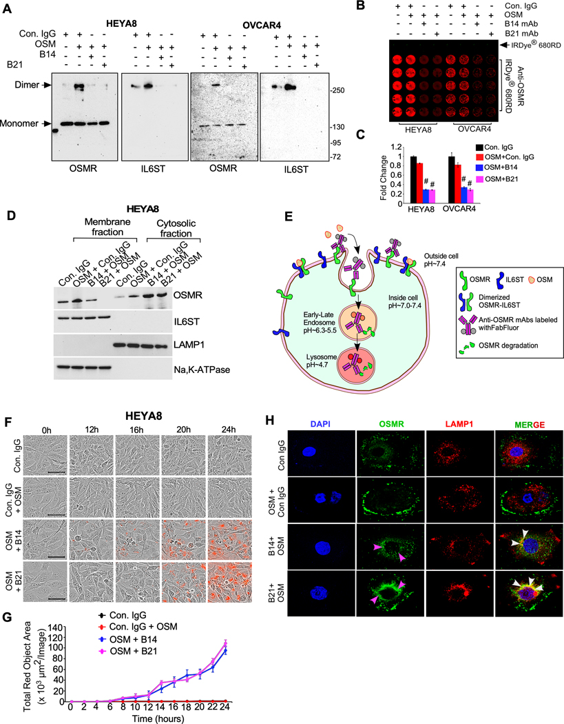
Figure. 5. Anti-OSMR antibodies abrogate heterodimerization of OSMR with IL6ST and induce internalization and degradation of OSMR.
A, HEYA8 and OVCAR4 cells were treated with B14 or B21 mAbs for 4h, then stimulated with OSM (100ng/mL) for 1h. Cell lysates were prepared after crosslinking with BS3 reagent and immunoprecipitated (IP) using anti-OSMR antibody, then resolved on SDS/PAGE. Dimers and monomers (arrows) were then detected using an anti-OSMR and anti-IL6ST antibodies. B, In-cell western blotting of OSMR in HEYA8 and OVCAR4 cells were treated with Control IgG, B14 and B21 mAbs in the presence of OSM for 24h. Primary antibodies bound on the cells were then labelled using secondary antibody 680RD infra-red dye and photographed. C, Each experiment was carried out in 5 biological and 3 technical replicates and fluorescent signals from (b) were quantified. D, HEYA8 cells were treated with B14 and B21 mAbs in the presence of OSM (100ng/mL) for 16h, then membrane and cytoplasmic fractions of cells were prepared, and Western blot was performed. LAMP1 and Na,K-ATPAse were used as internal controls of cytosolic and membrane fractions respectively. E, A schema depicts the process of anti-OSMR antibody mediated receptor internalization and degradation in cancer cells. Control IgG or B14 or B21 mAbs were labeled with Incucyte® Human FabFluor-pH Red Antibody Labeling Reagent and then added to cells along with OSM (100 ng/mL) and incubated for 24h. Red fluorogenic signals released due to the low-acidic pH when the FabFluor-labelled anti-OSMR antibody internalized to lysosomes were quantitated. F, HEYA8 cells were plated as described in (E) and time lapse imaging were performed to detect the fluorescence. Scale bar, 50μm. G, Quantitative assessment of internalized OSMR antibody complex based on the total red dot area per image as evaluated using Incucyte S3 software. H, Representative images of OSMR internalization and colocalization with LAMP1 lysosomal marker confocal microscopy. OVCAR4 cells were treated with control IgG, B14 and B21 mAbs in the presence of OSM for 16h and fixed and stained with OSMR-Alexa Fluor 488 (green) and LAMP1-Alex fluor 546 (red). Nuclei were stained with DAPI (blue). Student’s t test (two tailed, unpaired) was performed to determine P-value. Data represent means ± SEM. #P≤ 0.0001.

