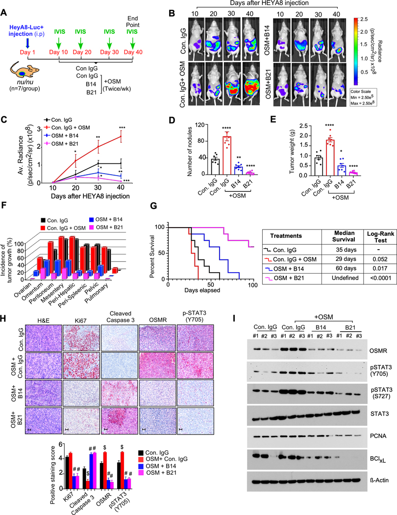
Figure. 6. Anti-OSMR antibodies reduced OSM mediated tumor growth and metastasis and improved overall survival rate of mice bearing ovarian cancer.
A, Schematic representation shows the experimental plan in athymic nude mice (nu/nu) injected with HEYA8-Luc+ cells intraperitoneally. Mice were treated with OSM (250 ng/kg body weight) 30 min after the injections of control IgG, B14, B21, antibodies (10mg/kg body weight) intraperitoneally for 5 weeks (N=7/ group) and then sacrificed. B-C, Representative images of tumor bearing mice were captured using an IVIS100 bioluminescence imager at the days indicated and the luminescence of tumor growth were quantitated. D-F, Number of tumor nodules, total tumor weight and incidence of tumor growth in specific organ sites were determined at the end of the experiment. G, Overall survival rate of ovarian cancer bearing nu/nu mice treated with control IgG, OSM, OSM+ B14 or OSM + B21 anti-OSMR antibodies twice per week for a period of 5 weeks (n=10/ group) and rate of survival was determined for a total of ~15 weeks (>100 days). Log-rank test was performed to determine P-value by comparing each group with control IgG group. H, Representative H&E-stained sections and IHC of indicated proteins in tumor tissues, that were isolated from mice in ‘B’ at the end of the experiment. Scale bar, 50 μm. i Western blots showing the expression of indicated proteins from three representative tumor tissues that were isolated from ‘a’ at the end of the experiment. Data represent means ± SEM. Student’s t test (two tailed, unpaired) and Dunnett’s multiple comparison tests were performed to determine P-Value in C, D, E and H. ****P≤ 0.0001, ***P≤0.001, and *P≤0.05, #P≤ 0.0001, $≤ 0.0001

