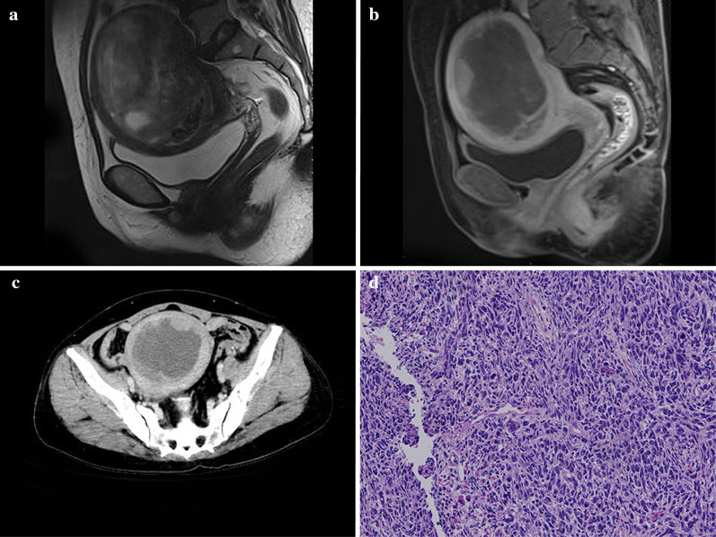
Fig. 2.
A 53-year-old woman with uterine carcinosarcoma (UCS). Sagittal T2WI magnetic resonance imaging (MRI) (a) showed a mixed signal tumor in the cavity complicated with intratumoral hemorrhage. Sagittal delayed enhanced T1WI (b) and axial computed tomography (CT) images in the venous phase (c) showed the marginal solid tissue component polyp-like enhancement with a central cystic component. The pathological image (d) showed uterine carcinosarcoma

