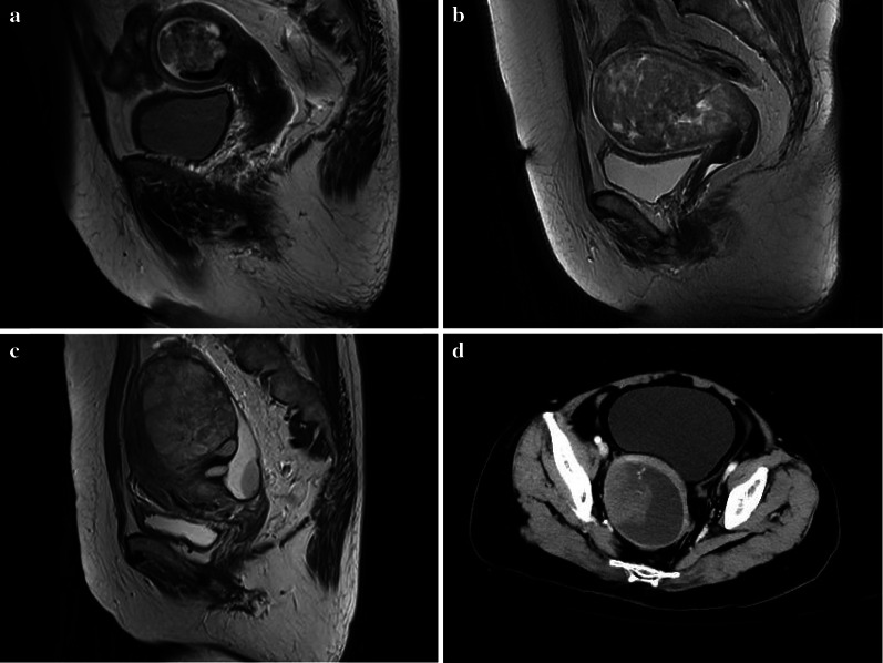
Fig. 4.
Images of uterine carcinosarcoma (UCS). Sagittal T2WI magnetic resonance imaging (MRI) of a 64-year-old woman (a) showed a mixed signal tumor in the cavity with multiple small round or irregular cystic areas in the internal tumor. Sagittal T2WI MRI of a 66-year-old woman (b) showed a mixed signal tumor in the cavity with many gap-like cystic areas. Sagittal T2WI MRI of a 49-year-old woman (c) showed a high signal tumor in the cavity with many oval cystic areas. Axial computed tomography (CT) images in the venous phase (d) of a 66-year-old woman showed a tumor mainly composed of cystic components. The tissue components showed nipple-like enhancement with a marginal cystic component

