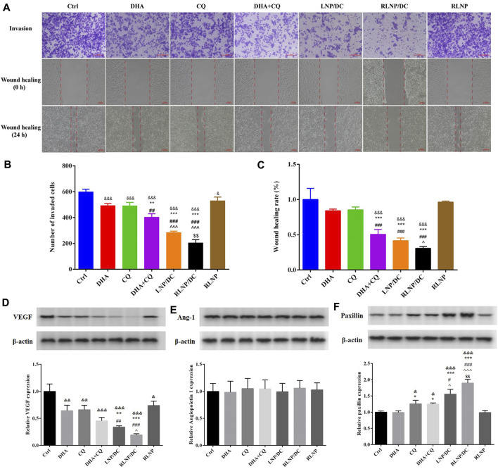
FIGURE 6.
Assessment of in vitro anti-metastasis effect and detection of metastasis-related proteins. (A–C) Transwell invasion assay and wound healing rate assay in HCT116 cells after treatment by DHA, CQ, DHA + CQ, LNP/DC, RLNP/DC, and RLNP. (D–F) Expression level of metastasis-related proteins including VEGF, Ang-1, and Paxillin after 24 h of treatment. Each bar represents the mean ± SD (n = 3). & p < 0.05, && p < 0.01, and &&& p < 0.001 vs. Ctrl; * p < 0.05, ** p < 0.01, and *** p < 0.001 vs. DHA; ## p < 0.01 and ### p < 0.001 vs. CQ; ^ p < 0.05 and ^^ ^ ^ p < 0.001 vs. DHA + CQ; $$ p < 0.01 vs. LNP/DC.

