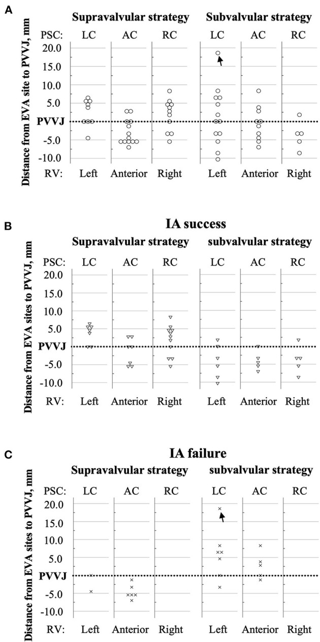
Figure 4.

Distances from the EVA sites to the PVVJ. The RVOT was arbitrarily divided by STJ, PVVJ, and longitudinal extensions of PSC commissures. The dotted line denotes PVVJ. The markers that coincided with the PVVJ located at the base of PSCs. The y-axis represents the distance from each EVA site to PVVJ. (A) Circles denote the EVA sites in total patients. (B) The triangles denote the EVA sites in the patients who had IA success. (C) The crosses denote the EVA sites in the patients who had IA failure. The black arrow marks the EVA site in PA. AC, anterior cusp; EVA, earliest ventricular activating; LC, left cusp; PA, pulmonary artery; PSC, pulmonary sinus cusp; PVVJ, pulmonary valve–ventricle junction; RC, right cusp; RV, right ventricle; RVOT, right ventricular outflow tract; STJ, sinotubular junction.
