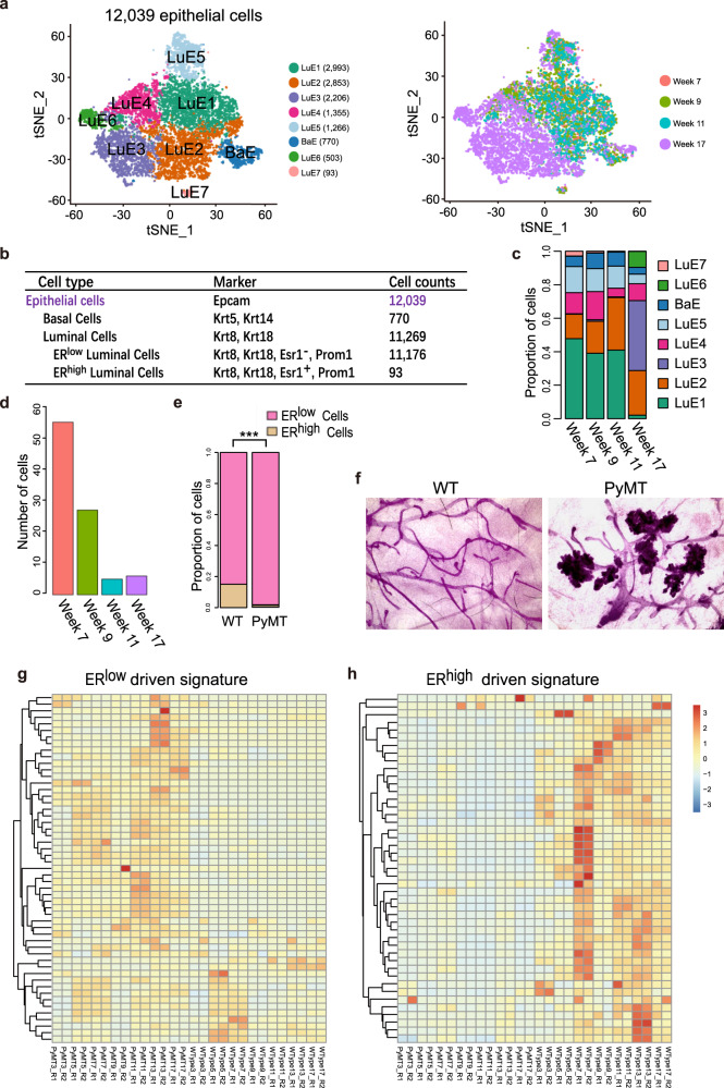
Fig. 2. Tumor cells originated from ERlow luminal cells during tumorigenesis.
a The t-SNE plot of 12,039 epithelial cells from the MMTV-PyMT mammary glands colored by cell types (left panel) and stages (right panel), week 7 (hyperplasia), 1,930 cells; week 9 (Adenoma/MIN), 2,886 cells; week 11 (early carcinoma), 2,038 cells; week 17 (late carcinoma), 5,185 cells. LuE1, 2, 3, 4, 5, 6, and 7: luminal epithelial cluster 1, 2, 3, 4, 5, 6, and 7; BaE: basal epithelial cluster. b Summary of cell counts and markers used for the identification of epithelial cell subsets from the mouse mammary glands and tumors. c The proportion of epithelial cell sub-clusters in different stages of tumor progression. d The number of ERhigh epithelial cells across different stages of tumor progression dramatically decreased. e The ratio of ERlow and ERhigh luminal epithelial cells in the mammary glands of MMTV-PyMT mice increased when compared to wild-type mice (WT), which suggests that the normal differentiation of ERhigh cell lineage was blocked, resulting in the accumulation of ERlow cancer cells. f Whole mounts of mammary glands from wild-type (WT) and MMTV-PyMT mice indicated that cancer cells mainly exist in location of alveolar cells. g Gene expression profile of ERhigh luminal signature was upregulated in epithelial cells from WT mice when comparing with epithelial cells from MMTV-PyMT mice. h ERlow luminal signature was highly expressed in MMTV-PyMT mouse epithelial cells relative to WT mice.

