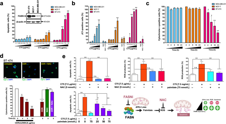
Fig. 2. FASN inhibition promotes high levels of apoptotic cell death in FASN-overexpressing breast cancer cells.
a. BT-474, MCF-7, MDA-MB-231 cells were treated with graded concentrations of C75 or vehicle (DMSO) for 48 h and apoptosis was evaluated by annexin V/propidium iodide staining using two-color flow cytometry. The inset shown the expression of FASN protein in the three cell lines. b Flow cytometry assessment of JC-1 fluorescence in cells treated with graded concentrations of C75 or vehicle (DMSO) for 48 h. Alternatively, cells were treated with 7.5 μg/mL C75 in the absence or presence of 5 mmol/L N-acetylcysteine (NAC). c Flow cytometry-based assessment of mitochondrial cytochrome c following exposure to C75 (7.5 μg/mL). d Top. Representative immunofluorescence images of cytochrome c staining in BT-474 cells treated with cerulenin in the absence or presence of N-acetylcysteine (NAC). Scale bar is 10 μm. Bottom. Flow cytometry-based assessment of mitochondrial cytochrome c following exposure to cerulenin in the absence or presence of NAC. e Left. Quantification of apoptosis and reactive oxygen species (ROS) in BT-474 cells treated with C75 in the absence or presence of NAC or palmitate. Right. Mitochondrial depolarization appears to be a common response to FASN inhibition irrespective of FASN status; however, the FASNi-driven decrease in ∆ψm levels appears to have to reach a certain threshold to elicit the release of mitochondrial cytochrome c accompanying apoptotic cell death, which is restricted to FASNi-sensitive cancer cells in an apparently ROS-dependent manner. All data are presented as mean ± SD (n = 3), p < 0.05 and p < 0.005 (* and **, respectively).

