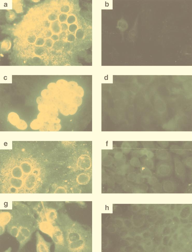
FIG. 3.
Immunofluorescence of SFV-2 infected cells. IFAs of uninfected and virus-infected M. dunni (a and b, respectively), Cf2Th (c and d, respectively), HeLa (e and f, respectively) and Vero (g and h, respectively) cells were done with plasma from animal Mn97 as described in Materials and Methods. Multinucleated syncytia as well as singly stained cells were seen in virus-infected cells, whereas no signal was seen in the uninfected cultures. Intense cytoplasmic and perinuclear staining was seen in infected M. dunni, HeLa, and Vero cells (a, e, and g, respectively). In Cf2Th cells, intense nuclear and diffuse cytoplasmic staining were seen (c).

