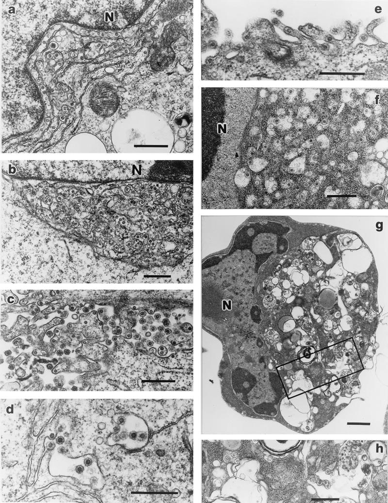
FIG. 4.
TEM of SFV-2-infected cells. SFV particles were seen intracellularly and budding from the plasma membrane. Infected M. dunni cells (a to c) had viral cores associated with extensively duplicated ER membranes, which were adjacent to the nucleus (a and b). Abundant mature particles were seen budding extracellularly from the plasma membrane (c). Infected Vero cells had few mature particles, which bud from the plasma membranes (d). In the case of infected Cf2Th cells (e and f) apoptotic cells were seen with few extracellular particles (e) and abundant accumulation of enveloped particles intracytoplasmically in dilated cisternae of the ER (f). In infected HeLa cells, enveloped particles were seen in the vacuolated cytoplasm of apoptotic cells (g and h). Few particles were seen to be budding from the plasma membrane. N, nucleus; the boxed region in panel g is magnified in panel h to show virus particles. Bars, 0.5 μm in panels a to f and h and 1 μm in panel g.

