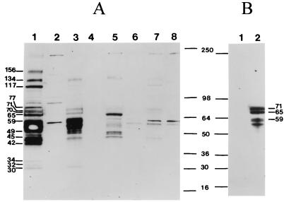
FIG. 5.
Western blot analysis of SFV-2-infected cells. Five micrograms of protein from lysates of the following SFV-2-infected cells (at a CPE of 2+) were immunoblotted with SFV-positive monkey plasma (A): M. dunni (lane 1), Cf2Th (lane 3), Vero (lane 5), and HeLa (lane 7). Uninfected cell lysates were the negative controls, as follows: M. dunni (lane 2), Cf2Th (lane 4), HeLa (lane 6), and Vero (lane 8) cells. In addition, Cf2Th cells were analyzed when the CPE was 3+ (B). Lane 1, uninfected cells; lane 2, infected cells. The sizes of some of the prominently visible SFV-2 proteins in M. dunni are indicated. The molecular masses were calculated from standard markers (SeeBlue; Novex, San Diego, Calif.) and are indicated in kilodaltons. The filter shown in panel A was incubated in substrate for 3 min and remained at room temperature for about 30 min before autoradiography. It was then exposed to X-ray film for 1 min. The filter shown in panel B was incubated in substrate for 10 s and immediately exposed to X-ray film for 10 s.

