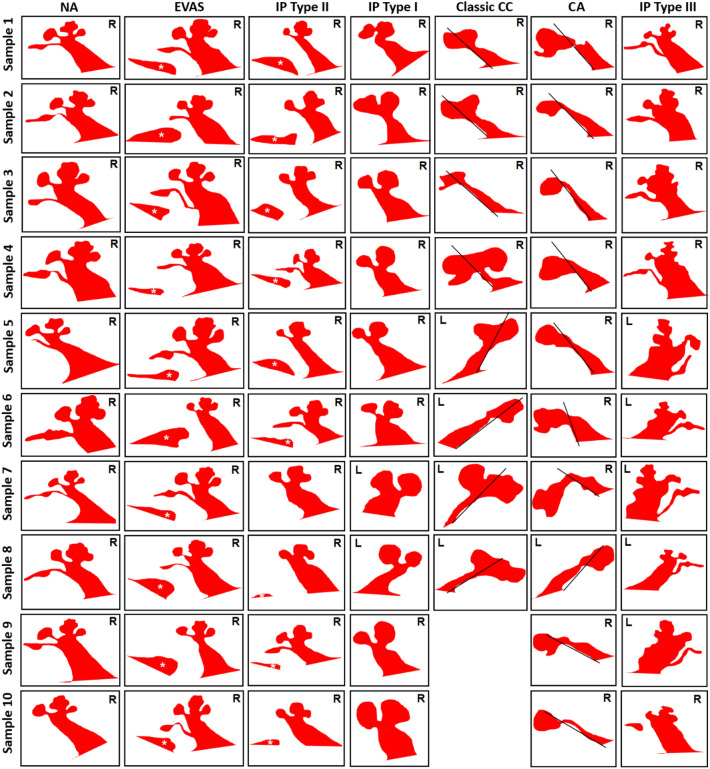
Figure 9.
Atlas of outer contours of mid-modiolar section of inner ear of different anatomical types. Classic CC has eight samples whereas other inner ear malformation types have ten samples. White asterisk refers to the vestibular aqueduct and the black line separates the cochlear and the vestibular portion in classic common cavity (CC) and shows the presence of vestibular cavity in cochlear aplasia (CA) type malformations. The white asterisk points to the enlarged VA. Normal anatomy: NA, Enlarged vestibular aqueduct syndrome: EVAS, incomplete partition: (IP) type I, II and III, Cochlear hypoplasia: CH. Images are not to scale. R: right ear; L: left ear.

