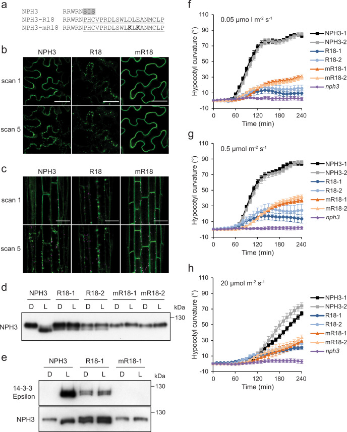
Fig. 7. Analysis of a constitutive 14-3-3 binding NPH3 variant.
a Amino-acid sequence of the NPH3-R18 and mR18 constructs. Residues 744–746 of NPH3 (grey shaded) were replaced with the R18 peptide sequence (underlined). Two lysine residues (bold) were introduced into the mR18 sequence to abolish 14-3-3 binding. b, c Confocal images of GFP-NPH3 (NPH3), GFP-NPH3 containing the R18 peptide sequence (R18) or the mutated R18 peptide sequence (mR18) (b) transiently expressed in leaves of N. benthamiana plants, dark-adapted before confocal observation and (c) in hypocotyl cells of etiolated transgenic Arabidopsis nph3 seedlings. Images were acquired immediately (scan 1) and after repeat scanning with the 488 nm laser (scan 5). GFP is shown in green and autofluorescence in magenta. Bar, 50 µm. These experiments were repeated at least twice with similar results. d Immunoblot analysis of total protein extracts from etiolated nph3 seedlings expressing NPH3, R18 or mR18 maintained in darkness (D) or irradiated with 20 µmol m−2 s−1 blue light for 15 min (L). Protein extracts were probed with anti-NPH3 antibodies. This experiment was repeated twice with similar results. e Far-western blot analysis of anti-GFP immunoprecipitations from etiolated nph3 seedlings expressing NPH3, R18 or mR18 maintained in darkness (D) or irradiated with 20 µmol m−2 s−1 of blue light for 15 min blue light (L). GST-tagged 14-3-3 isoform Epsilon was used as the probe. Blots were probed with anti-NPH3 antibody as a loading control (bottom panel). Phototropism of etiolated nph3 seedlings expressing GFP-NPH3 (NPH3), R18 or mR18 and nph3 mutant seedling irradiated with f 0.05 µmol m−2 s−1, g 0.5 µmol m−2 s−1 or h 20 µmol m−2 s−1 of unilateral blue light. Hypocotyl curvatures were measured every 10 min for 4 h, and each value is the mean ± SE of 18–20 seedlings from two independent biological replicates. Exact n values are provided in the Source data file.

