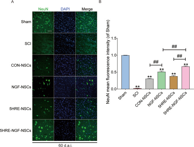
Fig. 6. Implement of NSCs and NGF, and application of hypoxia condition increased the expression of NeuN+ proteins at the day of 60.
A Immunofluorescence staining of NeuN in spinal cord lesions. The nuclei are labeled by DAPI (blue). The bright green dots are NeuN+ proteins, magnification was ×20. B Analysis of positive cells in immunofluorescence staining. ** signify P < 0.01 versus the Sham group, ## signify P < 0.01. Data are the mean values ± SD, n = 6.

