FIG. 4.
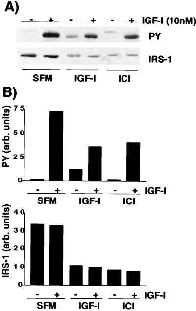
IGF-I-mediated degradation of IRS-1 results in reduced amounts of total tyrosine-phosphorylated IRS-1 following IGF-I stimulation. (A) MCF-7 cells were incubated in SFM for 24 h and then in SFM, or SFM supplemented with IGF-I (5 nM) or ICI 182780 (1 μM) for a further 24 h. Cells were or were not stimulated with IGF-I (10 nM) for 15 min and then lysed, and 50 μg of the resultant protein was separated by SDS–8% PAGE and immunoblotted with antiphosphotyrosine (PY) or anti-IRS-1 (IRS1) antibodies. (B) Densitometric analysis of gels shown in panel A. Gels were scanned and analyzed by using NIH Image 6.0. Values are presented as arbitrary (arb.) densitometric units. The top graph presents values for phosphotyrosine, and the bottom graph shows values for IRS-1.

