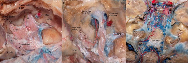
Figure 2.
(A) The dura of the lateral wall of the right CS has been removed to show the terminal branches of the ILT providing blood supply to CN III,IV as well asV1,V2,V3 branches of the trigeminal nerve. ILT giving off the superior branch, the artery of foramen rotundum, and the foramen ovale branch. (B) Another view from the same specimen after careful drilling of the foramen ovale showing the anastomotic network between the terminal branches of ILT and branches from the internal maxillary artery. Note the branches arising from MMA supplying the V3 segment of the trigeminal nerve. (C) Posterior view of the clivus in a colored silicone-injected human cadaveric specimen showing a rich anastomotic network between the dorsal meningeal branches arising from MHT and the jugular branches arising from neuromeningeal trunk, which is a branch of the ascending pharyngeal artery. APA, ascending pharyngeal artery; MMA, medial meningeal artery.

