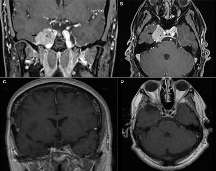
Figure 5.
Preoperative coronal (A) and axial (B) brain MRI with contrast showing heterogeneous enhancing tumor involving the right cavernous sinus (arrow). The patient is known to have right cavernous sinus meningioma for which he had stereotactic radiosurgery 4 years prior to presentation. Postoperative coronal (C) and axial (D) brain MRI with contrast showing gross total excision of the meningioma.

