Fig. 1.
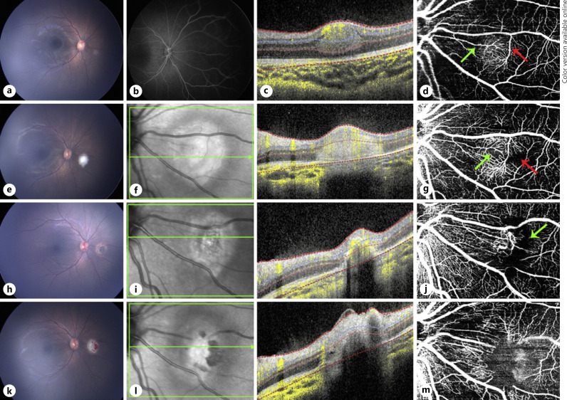
Multimodal image of case 1. a Color fundus photograph of a treatment-naive retinoblastoma lesion in the right eye. b FA only revealed mild hyperfluorescence at lesion area. c OCT B-scan showing a well-circumscribed hyper-reflective lesion involving retinal mid-layers. d En face 20 × 20° OCTA revealed the feeder and draining dilated vessels (green arrow and red arrow, respectively) and dense intrinsic vascularity. e Color fundus photograph of the same eye immediately after the first TTT. f OCT B-scan of the same lesion immediately after the first TTT shows an enlargement, not definite margins, and an increase in hyper-reflectivity compared to pre-TTT. g En face 20 × 20° OCTA immediately after first TTT does not show major vascular differences apart from an increase in vessel reflectivity and less perfused areas (green and red arrows). h Color fundus photograph 1 month after the first TTT of the same tumor. i OCT B-scan shows shrinkage of the lesion with indistinct borders. j En face 20 × 20° OCTA 1 month after the first TTT treatment revealed a drastic decrease in intrinsic vessel network density replaced with a dark area (green arrow), with retraction of the draining and feeder vessels and traction of surrounding vessels from the treated lesion. k−m correspond to images immediately after second TTT, showing a similar effect obtained immediately after the first TTT treatment was done. m En face 20 × 20° OCTA obtained immediately after the second TTT revealed minimum tumor intrinsic vascular network replaced by a dark mild hyper-reflective area due to inflammation caused by treatment. FA, fluorescein angiography; OCTA, optical coherence tomography angiography; TTT, transpupillary thermotherapy.

