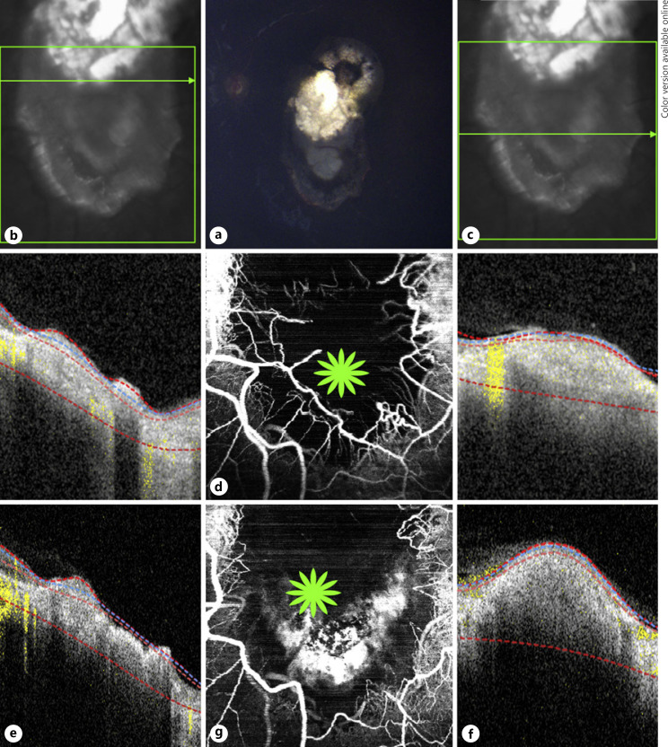
Fig. 3.
Multimodal image of case 3. a Color fundus photograph of a retinoblastoma lesion with type 3 regression and a tumor recurrence at the inferior aspect of the lesion in the left eye. OCT B-scans (b) over the superior portion showing a disorganized retinal atrophic area and another cut (c) over the tumor relapse depicting an elevated hyper-reflective lesion. d En face 20 × 20° OCTA shows a network of dilated tortuous medium-sized vessels at the borders of tumor relapse and a dark area at the center revealing absence of vessels. OCT B-scans over the same areas immediately after the third TTT session with the superior portion (e) still having an atrophic appearance and over the relapse area (f), an increase in size and reflectivity compared to pre-TTT (c) due to tissue inflammation caused by the treatment. g En face 20 × 20° OCTA over the same area immediately after the third TTT shows the dilated vessels surrounding the relapse are not present and next to that area a hyper-reflective signal (green star) caused by the treatment inflammation. OCTA, optical coherence tomography angiography; TTT, transpupillary thermotherapy.

