Fig. 5.
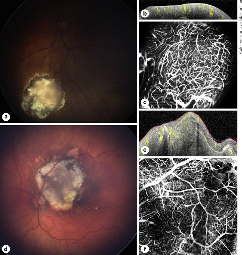
Multimodal image of cases 5 and 6. a Color fundus photograph of a retinoblastoma lesion from case 5 with type 3 regression in the left eye, 1 month after the fourth TTT session. b OCT B-scan over the tumor shows an elevated vascularized hyper-reflective lesion involving all retinal layers. c En face 20 × 20° OCTA of the lesion shows a network of tortuous and dilated medium- and small-sized vessels. d Color fundus photograph of a retinoblastoma lesion from case 6 with type 3 regression with a cystic area at the temporal superior border in the left eye, 1 month after the third TTT session. e OCT B-scan over the lesion showing an elevated hyper-reflective lesion with an irregular anterior border and intralesional calcifications causing shadowing. f En face 20 × 20° OCTA of the same lesion shows a disorganized network of tortuous dilated and large-, medium-, and small-sized vessels and a dark area at the superotemporal border caused by the cystic portion and intralesional calcifications. OCTA, optical coherence tomography angiography; TTT, transpupillary thermotherapy.

